
Brain Scans Offer New Insights into Migraine Mysteries
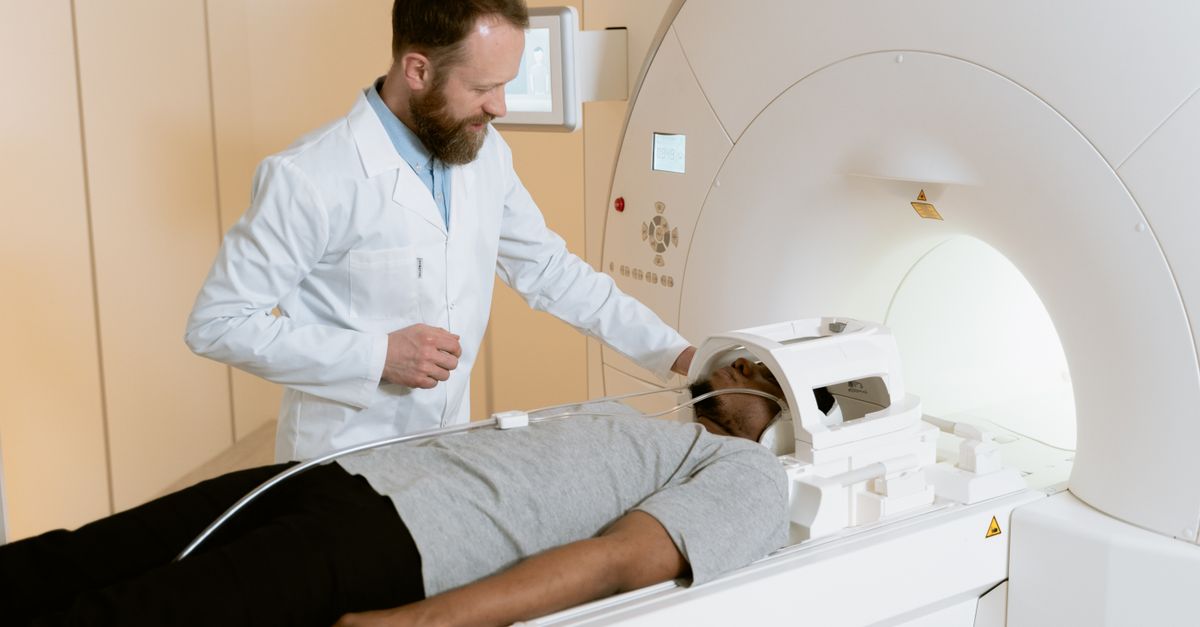
Advanced brain imaging is unraveling the complexities of migraines, offering fresh perspectives on diagnosis and treatment.
The debilitating pain of migraines affects over a billion people globally, often leaving sufferers desperate for relief. While the exact cause of migraines remains elusive, recent advancements in brain imaging are opening new pathways to understanding this complex neurological condition. By visualizing the brain activity of migraine sufferers, researchers are uncovering patterns that could revolutionize both diagnosis and treatment.
The Role of Advanced Imaging Techniques
MRI and PET scans have long been staples in medical diagnostics, but their application in migraine research is relatively novel. These imaging techniques allow doctors to observe changes in brain function and structure that occur during a migraine attack. For instance, functional MRI (fMRI) can detect alterations in blood flow and neural activity, providing a window into the brain's response to migraine triggers.
Studies at institutions like the University of California, Los Angeles, are leveraging these technologies to identify biomarkers associated with migraines. Researchers have observed distinctive patterns of brain activity in areas responsible for processing pain and sensory information. These findings not only enhance our understanding of migraines but also challenge the traditional notion that they are solely vascular events.
What the Scans Are Revealing
One of the most intriguing discoveries from brain scans is the role of the hypothalamus, a small region deep in the brain often implicated in hormone regulation and autonomic functions. Imaging studies have shown that the hypothalamus may become hyperactive before and during a migraine, suggesting it could be a key player in the onset of attacks.
Additionally, scans have revealed that migraines might involve a broader network of brain regions than previously thought. This includes the brainstem and areas linked to processing emotional and sensory stimuli, contributing to the diverse symptoms experienced by migraine sufferers, such as light sensitivity and mood changes.
Implications for Diagnosis and Treatment
The insights gained from brain imaging are not just academic. They hold real potential for improving clinical outcomes. For one, identifying specific brain activity patterns could lead to more accurate and earlier diagnosis. This is particularly significant in differentiating migraines from other types of headaches, which can guide more effective treatment plans.
Treatment options could also become more personalized. As researchers pinpoint which brain regions are most active during migraines, they can develop targeted therapies that address those specific areas. This could include neurostimulation techniques that modulate brain activity without medication, a promising avenue for patients who do not respond well to conventional treatments.
Challenges and Future Directions
Despite these promising developments, challenges remain. Brain imaging is expensive and not yet feasible as a routine diagnostic tool for migraines. Moreover, the interpretation of complex brain scans requires specialized expertise, which can limit accessibility.
Nonetheless, the future looks promising. As technology advances, costs are likely to decrease, making these scans more accessible to a wider population. The continued integration of artificial intelligence in analyzing brain imaging data could also streamline the process, offering quicker and more precise interpretations.
Dr. Emily Zhang, a neurologist at the Mayo Clinic, emphasizes, "We are just scratching the surface of what brain imaging can tell us about migraines. The goal is to refine these techniques to the point where they can be used routinely to not only diagnose but also predict and prevent migraine attacks."